

Right leg
Left leg instructions
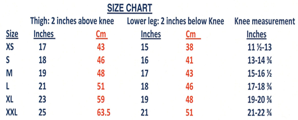
Size chart^
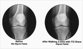
X Ray alignment of bones of knee joint behind the patella after just 2 minutes walking while wearing In the Groove Knee Re-trainer. Patella is highlighted.
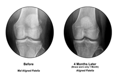
Xray before and after wearing Knee re-trainer for 1 month, 3 months later. Patella still in the groove.
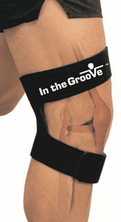
Knee Re-trainer Brace
Knee Re-trainer Brace is a comfortable wrap around re-trainer brace that does a modified physical therapy exercise while you wear it during daily activity. Best for side-to-side knee pain like PFS, runner’s knee, skier’s knee, jumper’s knee, etc. Get in the Groove to keep on the move
Have knee pain? Re-Train muscles of the knee joint to:
- reduce pain
- improve mobility
- feel more stable
- slow further damage to bones of the knee joint^
Know before you buy technology with a simple two-handed maneuver you can do on yourself. right leg ^ left leg^
Small Business • Made in USA • Woman owned •Patented
59.95 shipping within lower 48 by USPS Priority included, call (517) 781-6030 or email abrown@inthegroovebrace.com for other options or additional cost.
Not returnable if worn on skin or if not in re-sellable condition. Do maneuver to determine effectiveness for you. Know before you buy. Limited 30-day warranty.
-8% for Amazon day
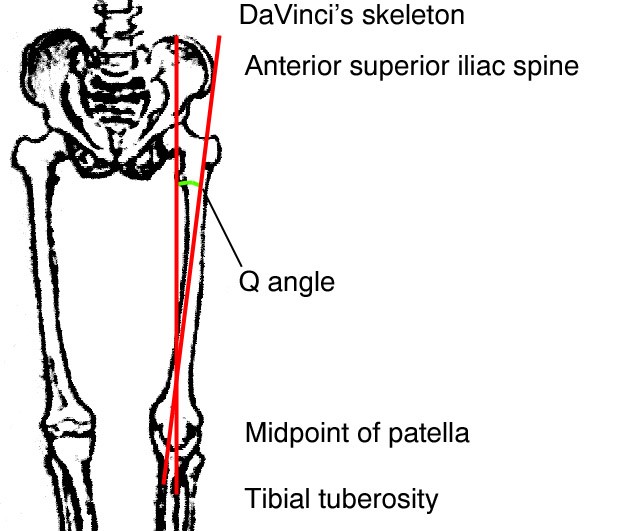
Q 4. Angle Diagram and increased incidence of PFS in Women
As the angle between the hip crest and the tibial tuberosity increases, the femur is unable to sit within the menisci wells of the tibial plateau. Out of the wells, the femoral condyles (or ends) crush the menisci wells and damage the tissues of the knee joint.
Women naturally have a greater Q angle than men and they aggravate it by doing the model walk where one foot is placed directly over the other foot rather than pointing straight ahead. This explains why women are more prone to patellofemoral syndrome.

How Does It Work
Game Changer Brace
A Game Changer Brace Helps in Improving Walking, Jogging and Workout Exercise, The GAMECHANGER restores natural tracking and thus restores normal movement. When normal movement is restored, muscles are able to rehabilitate or train themselves to maintain better alignment and thrush the brace is no longer needed after 2 or 3 months, unless there is already severe damage to knee joint tissues. If there is severe damage or arthritic changes, then the brace may be needed long term.
HOW DOES IN THE GROOVE KNEE RE-TRAINER BRACE™ WORK?
KNEE JOINT FACTS and ANATOMY
The Knee joint is the largest joint in the body and is more likely to be injured than is any other joint in the body.
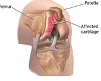
Anatomy of the knee joint showing the femur, thigh bone, patella or knee cap and how the patella tracks over the femur end which looks like an upside-down V.
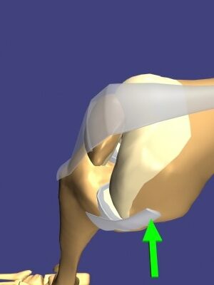
This is a picture of the knee bent showing the tendon on the side of the knee, green arrow points to it, and the large tendon that holds the patella. The patella moves over and in the groove of the femur. If a tight sleeve brace is worn, it will force the patella closer to the femur and if they touch, direct physical contact can damage to the bone ends causing pain with every movement of the knee joint. This is patellofemoral syndrome or PFS. The patella and femur damage each other when they scrape over eachother.
The green arrow points to medical collateral ligament
which often gets streched causing instability of knee joint.
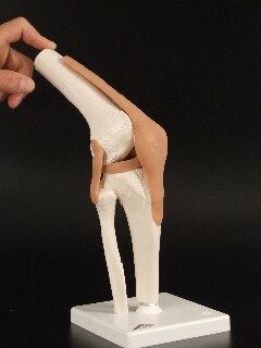
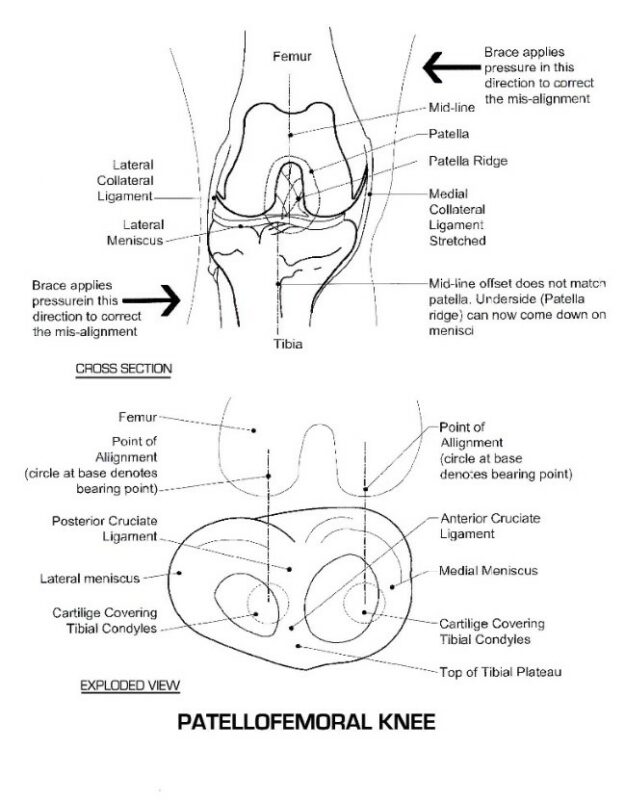
This is the model of the knee bent. You can see how the patella is suspended in the tendon and moves over the femur with movement of the knee joint. If the bones are pressed close together, they end up scraping each other and this causes pain and may damage the boney surfaces that grind over each other. In the Groove Knee Re-Trainer Brace™ has stays that apply gentle pressure to the thigh and lower leg to align the bones so the patella can track “in the groove” of the femur/thigh.
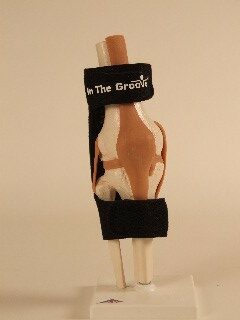
This is a model of the knee joint with the tendons attached as beige and the bones are white. In the Groove Knee Re-Trainer Brace™ is worn with the longest strap above the knee joint, body of the brace behind the knee joint and the shorter strap below the knee joint.
The In the Groove™ Knee Re-training Brace™ helps to align the thigh over the lower leg which supports the entire weight of the body. Any brace that does not align the thigh over the lower leg (tibia) does not help to reduce the damage done to the knee joint bones and cartilage with each and every movement of the knee.
When normal alignment is restored, pain is reduced, more normal function is restored and there is the potential to not only reduce further damage to the knee joint, but also to allow the body to heal where possible.
In fact, after 2 or 3 months of use, most people find that they no longer need the In the Groove™ Knee Re-training Brace™, unless there has already been severe damage to knee joint tissues or arthritic changes, then it may be needed long term.
How Do I Use In the Groove Knee Re-Training Brace™
1. Wrap the In the Groove Knee Re-Training Brace™ around the sore knee with the body of the brace on the back of the knee
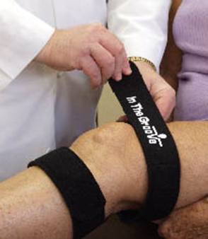
2. Attach the top (longer) strap around the thigh and attach the hook section to the loop strap so that it is comfortable and tight enough to stay on your leg when you stand up. Do not pull the strap so tight that it cuts circulation.
3. Now attach the lower (shorter) strap the opposite direction so that the hook section attaches to the loop strap. Again, wrap it around the lower leg so that it is comfortable and tight enough to stay on the leg when you stand up, but not so tight that it cuts circulation. If you are going to be sitting for a long time, or on a long flight, you can either un-attach the lower strap or attach it looser to not reduce circulation and re-attach strap before you get up.
KNEE FACTS:
The knee is designed to support 400 POUNDS, but a squat puts 4X pressure on knee.
In a squat a 100-pound person is at the maximum weight for the knee.
The knee will NEVER WEAROUT out unless it is injured.
The KNEE joint is the LARGEST JOINT in the human body.
The knee joint can be DAMAGED:
- Traumatically, as in sports injury
- Chronically, when it structurally compensates for ankle malalignment by mal-aligning the knee so that the leg is aligned when in motion as walking.
- Medical disease that the body recognizes “joint tissues as foreign invaders” causing the body’s own white cells to attack the tissues of the knee joint as in rheumatoid arthritis, lupus, etc.
- Structural deviations from normal knee joint as in genetic diseases such as dwarfism, gigantism, etc.
- Repetitive strain injury causes micro tears to the muscles and the tendons faster than tissue repair can occur. This weakens the structures around the patella and becomes less able to hold it in the correct position.
Knee anatomy:
Knee anatomy with parts labeled You can see how little room there is in the knee joint itself so if a piece of meniscus gets inside it will cause pain and limit motion of knee.
Top of this picture shows thigh bone ends that fit into meniscus wells of lower leg
View of knee joint looking at top of lower leg (bottom) with 2 wells formed by meniscus to hold thigh bone end. If meniscus is damaged knee joint becomes unstable.
Patellofemoral Pain is the most common knee disorder. There are two types:
- Patellofemoral instability patients have pain at the end range extension.
- Patellofemoral arthritic degeneration patients have pain with deep knee flexion, “closed chain” activities.
- Both groups should perform exercises in pain free range.
Women are more likely to have patellofemoral syndrome, PFS, then men. Men’s legs hang fairly straight from the hip but women’s legs bend towards each other. The greater Q angle women have is so that the pelvic bones can move more so that during delivery, the baby’s head has room to go through the opening. You can see that the knees do not hang straight, but their knees are slightly closer together. This is often accentuated by the “model walk”.
Women’s Q angle, as drawn in red, allows for a larger opening during childbirth. You can see that the knees do not hang straight, but their knees are slightly closer together. This closeness is often accentuated by the “model walk”.
If knee joint is MAL ALIGNED no matter the cause the knee cannot move correctly and damage to the individual elements of the knee joint results.
Mal tracking of the patella is often not detectable by the naked eye.
The exact cause of patellofemoral pain isn’t known. It probably has something to do with the way the patella moves on the groove of your thigh bone (femur).
If the knee were just a hinge joint you could not pivot your leg or foot.
A common misconception is that the patella only moves in up-and-down direction. In fact, it also tilts and rotates, so there are various points of contact between the undersurface of the patella and the femur.
Restoring normal structure of the knee joint reduces further damage to the knee joint and relieves the pain caused by tissue damage. When normal structure is restored and maintained, muscles can train and strengthen to help maintain the normal structure.
Wearing a knee brace that does not align the leg bones behind the patella.
Trains the muscles to maintain mal-alignment
Mal-alignment must be unlearned
Then relearned muscle memory to align the bones and maintain it
Knee braces and Sleeves are probably best reserved for use in patients with lateral subluxation that can be seen with the naked eye and can be easily palpated.
The use of any KNEE BRACE OR SLEEVE should NOT a substitute for THERAPEUTIC EXERCISES.
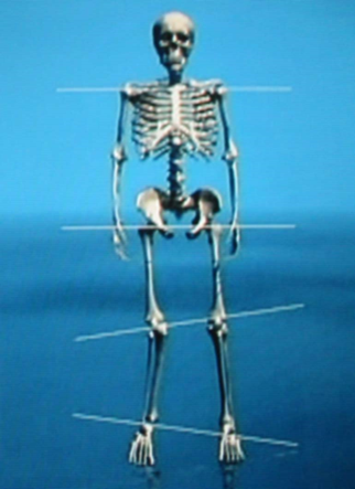
Remember EVERY JOINT affects other JOINTS in the body. The body wants to walk straight.
If one joint is out of alignment the joint above and below will also be out of alignment the body as a unit will look like it’s aligned.
There is a song that goes:
The foot bone is connected to the leg bone
The leg bone is connected to the thigh bone
Do the skeleton dance
The thigh bone is connected to the back bone
The back bone is connected to the neck bone.
TO SEE DANCING SKELETON CLICK:
https://youtu.be/Pbl4BNkAq_U
To Test your Right leg:
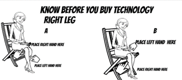
- Gently Apply pressure to the outside of the right knee just two inches above the knee with right hand, then put left hand about two inches below the right knee on the inside of the right leg.
- Now gently apply pressure toward the opposite hand and bend and kick the leg at the knee. If there is no pain and the muscles feel relaxed, then you need an A.
- If the muscles of the right knee tighten and there is pain, reverse hand positions so that the right hand is below the right knee on the outside of the leg and the left hand is above the knee on the inside of the leg. If this feels good or the muscles relax, you need a B.
- IF NEITHER WAY FEELS GOOD, DO NOT BUY THIS BRACE AS IT WILL NOT HELP YOU, UNLESS you are a cyclist who has knee pain only after cycling.
To Test your Left Leg:
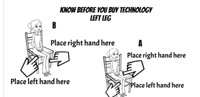
- Gently Apply pressure to the outside of the left knee just two inches above the knee with left hand, then put right hand about two inches below the left knee on the inside of the left leg.
- Now gently apply pressure toward the opposite hand and bend and kick the leg at the knee. If there is no pain and/or the muscles feel relaxed, then you need an B.
- If the muscles of the right knee tighten and/or there is pain, reverse hand positions so that the right hand is below the right knee on the outside of the leg and the left hand is above the knee on the inside of the leg. If this feels good or the muscles relax, you need an A.
- IF NEITHER WAY FEELS GOOD, DO NOT BUY THIS BRACE AS IT WILL NOT HELP YOU, UNLESS you are a cyclist who has knee pain only after cycling.